World Journal of Nephrology and Urology, ISSN 1927-1239 print, 1927-1247 online, Open Access
Article copyright, the authors; Journal compilation copyright, World J Nephrol Urol and Elmer Press Inc
Journal website http://www.wjnu.org

Case Report

Volume 7, Number 3-4, November 2018, pages 85-87


A Large Jackstone Calculus Incidentally Detected on CT Examination: A Case Report With Literature Review

Figures

Figure 1.
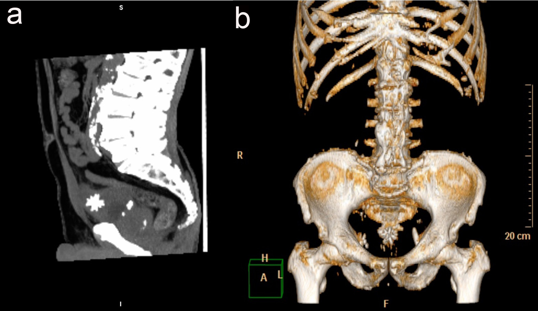
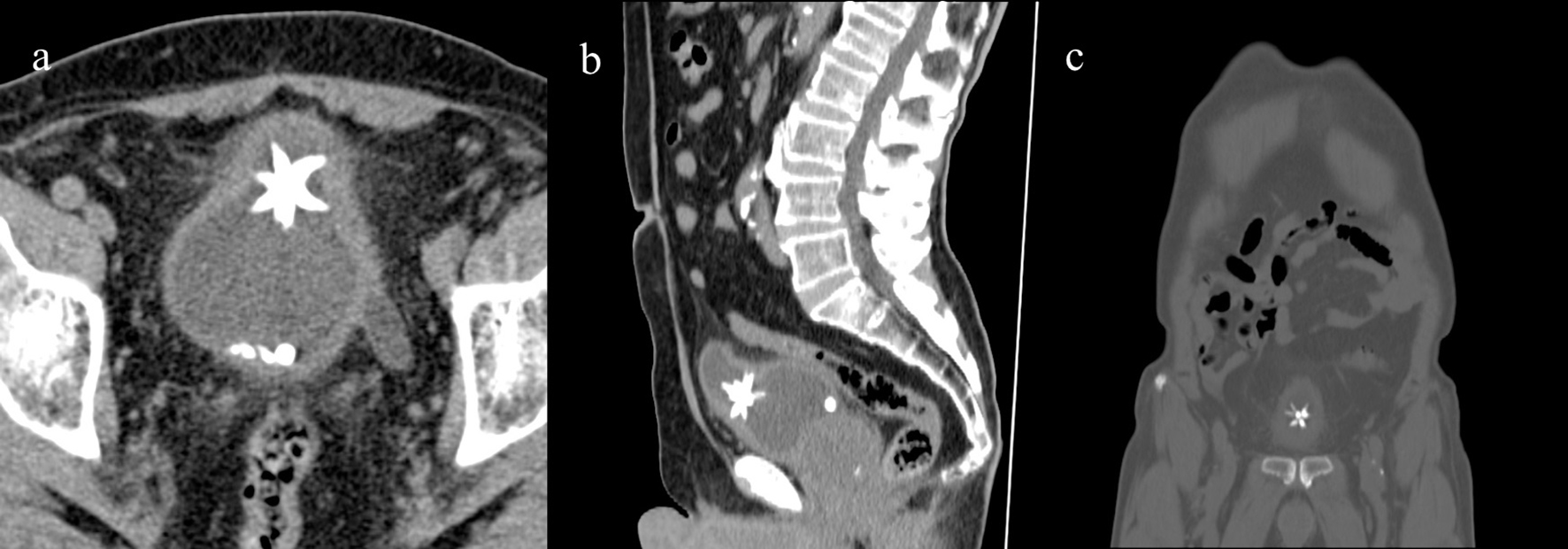
Figure 1. Axial CT showing the Jackstone calculus between the bladder dome and the body with adjacent wall thickness. Other calculi with regular appearances were also visible at the bladder base (a), MPR reconstruction on sagittal (b) and coronal plane (c).
Figure 2.
Figure 2. The Jackstone calculus on MIP 3D reconstruction on sagittal plane (a) and on VR (b).
Figure 3.
Figure 3. Picture of six-point Jack’s toys.