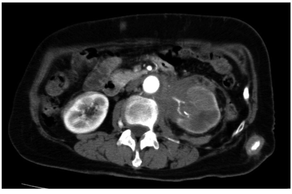
Figure 1. Computed tomography showed a mass around the left renal hilum and subcutaneous abdominal masses.
| World Journal of Nephrology and Urology, ISSN 1927-1239 print, 1927-1247 online, Open Access |
| Article copyright, the authors; Journal compilation copyright, World J Nephrol Urol and Elmer Press Inc |
| Journal website http://www.wjnu.org |
Case Report
Volume 1, Number 4-5, October 2012, pages 121-124
Cutaneous Metastases From Transitional Cell Carcinoma of the Renal Pelvis: A Case Report
Figures
Tables
| Case | Author | Sex | Age | Initial Treatment | Adjuvant Therapy | Duration between surgery and skin metastases (month) |
|---|---|---|---|---|---|---|
| 1 | Ando et al | male | 67 | nephroureterectomy | chemotherapy | 27 |
| 2 | Chitale et al | male | 68 | nephroureterectomy | none | 2 |
| 3 | Zirwas et al | male | 43 | nephroureterectomy | none | 48 |
| 4 | Lin et al | female | 68 | nephroureterectomy | chemotherapy + radiation therapy | 18 |
| 5 | our case | female | 73 | none | none | 0 |
| Case | Appearance of skin metastasis | Distribution of skin metastases | Other metastatic regions | Treatment | Outcome | Follow-up (month) |
|---|---|---|---|---|---|---|
| 1 | zosteriform | left chest wall | left axillary LN | chemotherapy + radiation therapy | alive | 10 |
| 2 | unknown | back, abdomen, limbs | lung, brain | none | dead | 1 |
| 3 | vascular-appearing nodule | shoulder | sacroiliac area, lung | chemotherapy | unknown | unknown |
| 4 | nodular | arm, abdomen | local, liver, left kidney | none | dead | 1 |
| 5 | nodular | abdomen | none | chemotherapy | dead | 3 |