World Journal of Nephrology and Urology, ISSN 1927-1239 print, 1927-1247 online, Open Access
Article copyright, the authors; Journal compilation copyright, World J Nephrol Urol and Elmer Press Inc
Journal website http://www.wjnu.org

Case Report

Volume 2, Number 2, November 2013, pages 82-84


Cystic Lymphangioma of the Adrenal Gland: Case Report

Figures

Figure 1.
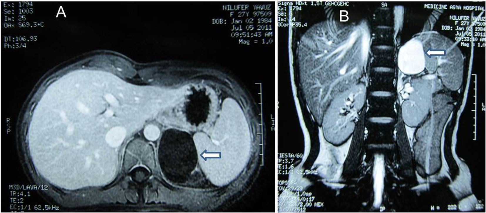
Figure 1. (A) MRI. Post-contrast axial T1-weighted image demonstrates cystic mass at left suprarenal location (arrow). (B) MRI. Post-contrast coronal breath hold T2-weighted image demonstrates cystic mass of 60 ´ 40 mm in diameter with homogenous high signal intensity (arrow).
Figure 2.
Figure 2. Histologically cystic lesion covered by flat endothelial cells adjacent to the normal-appearing adrenal gland.
Figure 3.
Figure 3. Flat endothelial cells are positive for D2-40 (A), and positive for CD31 (B).