World Journal of Nephrology and Urology, ISSN 1927-1239 print, 1927-1247 online, Open Access
Article copyright, the authors; Journal compilation copyright, World J Nephrol Urol and Elmer Press Inc
Journal website http://www.wjnu.org

Case Report

Volume 3, Number 1, March 2014, pages 49-53


The Heterogeneity and Controversy of C1q Nephropathy: A Report of Two Cases and Review of the Literature

Figures

Figure 1.
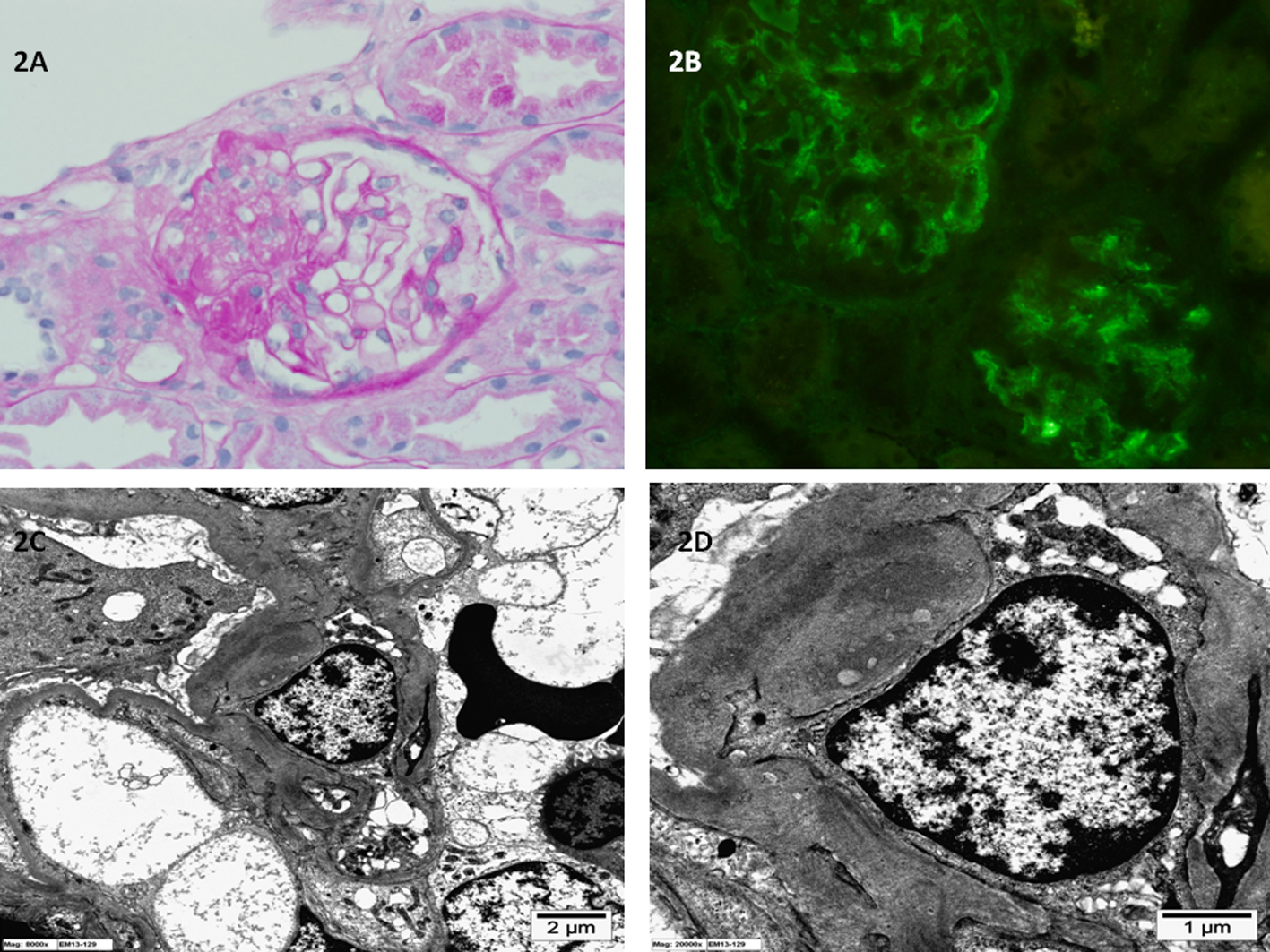
Figure 1. (A) Light microscopy of kidney biopsy specimen shows mild mesangial proliferation and mesangial matrix expansion (periodic acid-Schiff stain; original magnification × 400). (B) Immunofluorescence microscopy of kidney biopsy specimen shows global mesangial staining with complement C1q (anti-C1q antibody immunofluorescence; original magnification × 400). (C, D) Electron microscopy of kidney biopsy specimen shows electron dense immune deposits in the glomerular mesangium with extension into the paramesangium (uranyl acetate, lead citrate stain; original magnification × 8,000 and × 15,000 respectively).
Figure 2.
Figure 2. (A) Light microscopy of kidney biopsy specimen shows segmental sclerosis and the glomerular, basement membrane is of normal thickness (periodic acid-Schiff stain; original magnification × 400). (B) Immunofluorescence microscopy of kidney biopsy specimen shows global mesangial staining with complement C1q (anti-C1q antibody immunofluorescence; original magnification × 400). (C, D) Electron microscopy of kidney biopsy specimen shows electron dense immune deposits in the glomerular mesangium (uranyl acetate, lead citrate stain; original magnification × 8,000 and × 20,000 respectively).