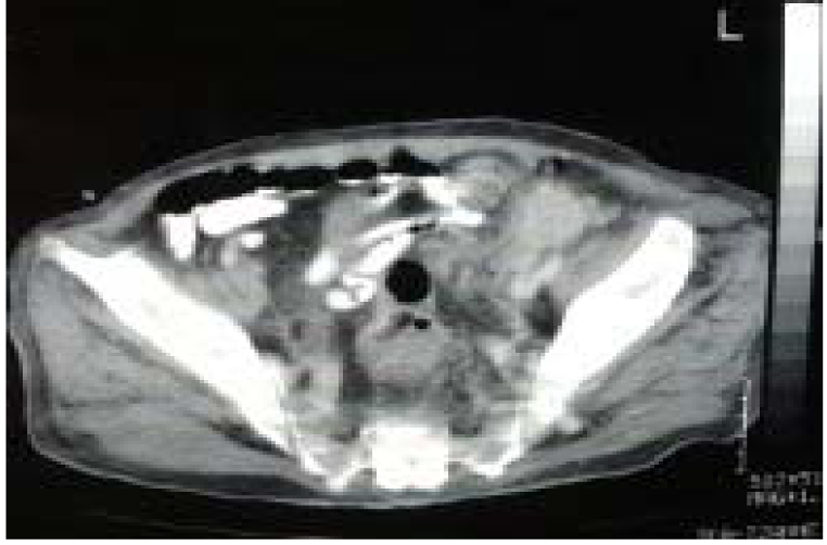
Figure 1. Computed tomography (CT) of the abdomen showed a 6 cm solid mass in the left groin.
| World Journal of Nephrology and Urology, ISSN 1927-1239 print, 1927-1247 online, Open Access |
| Article copyright, the authors; Journal compilation copyright, World J Nephrol Urol and Elmer Press Inc |
| Journal website http://www.wjnu.org |
Case Report
Volume 3, Number 1, March 2014, pages 58-62
Squamous Cell Carcinoma of the Urinary Bladder: A Clinicopathological Study of Four Cases and a Review of the Literature
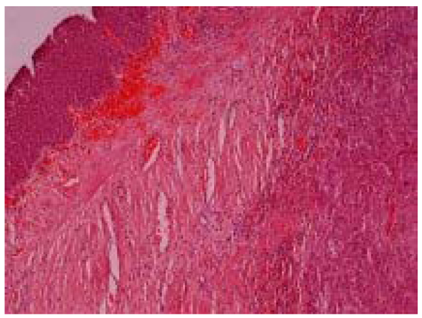
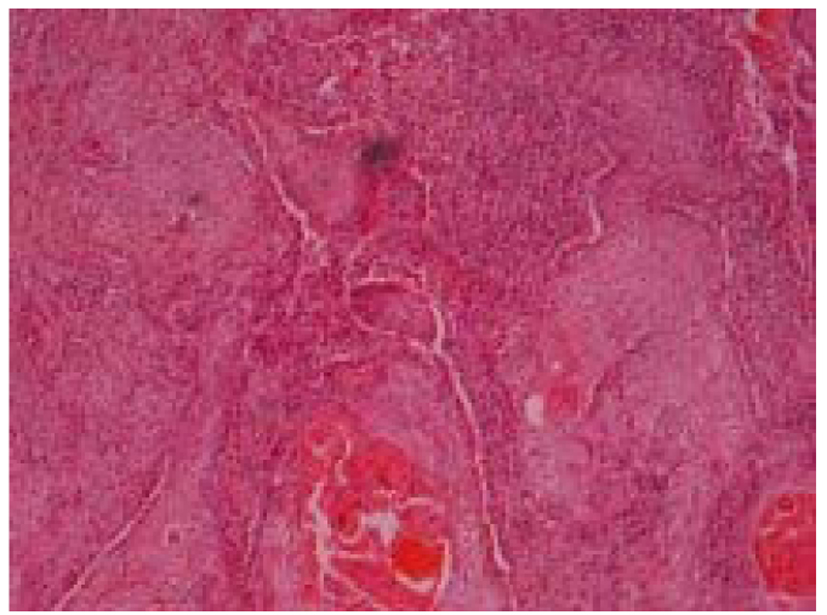
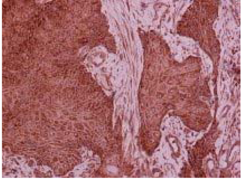
Figures