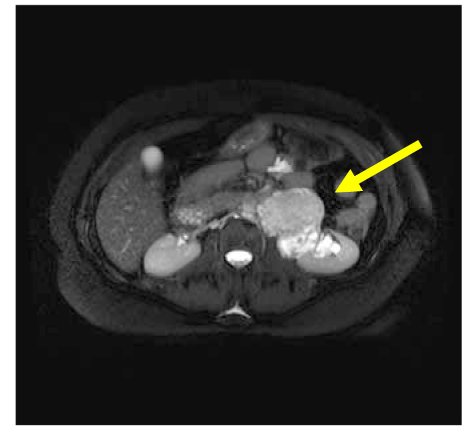
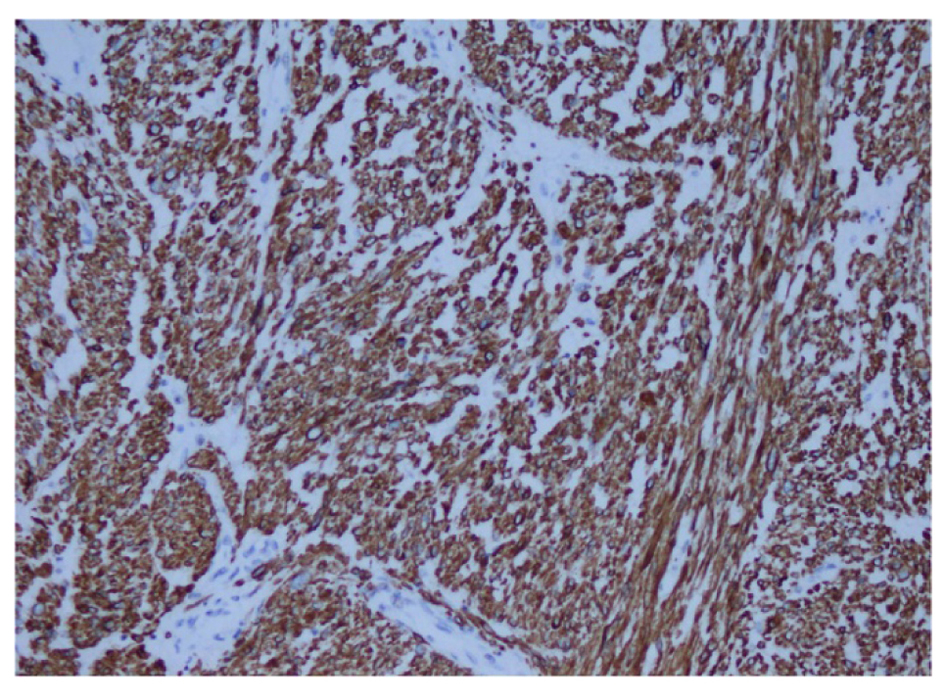
Figure 1. Magnetic resonance imaging: mass in pelvis renalis of left kidney (arrow), the coronal reconstruction shows.
| World Journal of Nephrology and Urology, ISSN 1927-1239 print, 1927-1247 online, Open Access |
| Article copyright, the authors; Journal compilation copyright, World J Nephrol Urol and Elmer Press Inc |
| Journal website http://www.wjnu.org |
Case Report
Volume 3, Number 1, March 2014, pages 63-65
High-Grade Primary Leiomyosarcoma of the Kidney
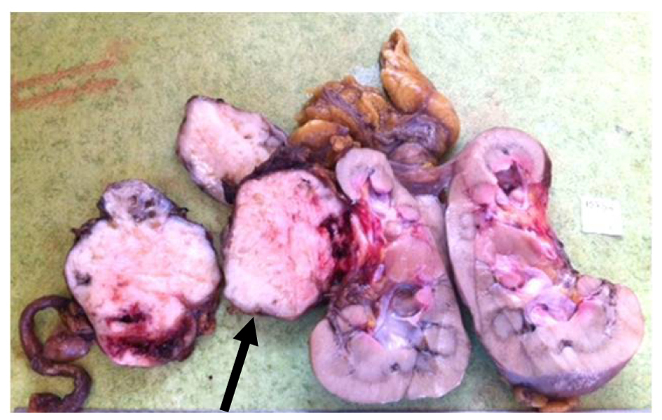
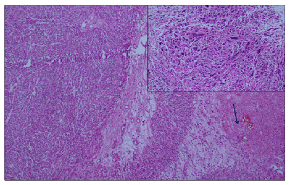
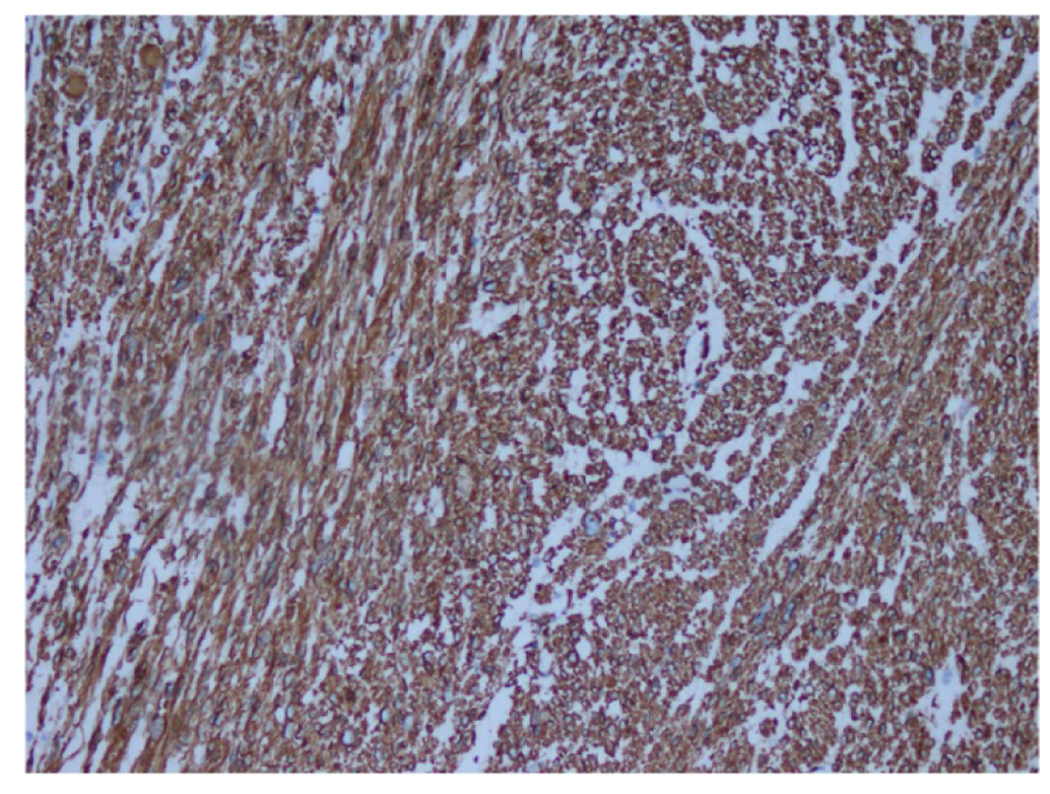
Figures