World Journal of Nephrology and Urology, ISSN 1927-1239 print, 1927-1247 online, Open Access
Article copyright, the authors; Journal compilation copyright, World J Nephrol Urol and Elmer Press Inc
Journal website http://www.wjnu.org

Case Report

Volume 3, Number 2, June 2014, pages 106-109


The Case of Pyelonephritis and Renal Cyst Infection Diagnosed by Diffusion-Weighted Magnetic Resonance Imaging

Figures

Figure 1.
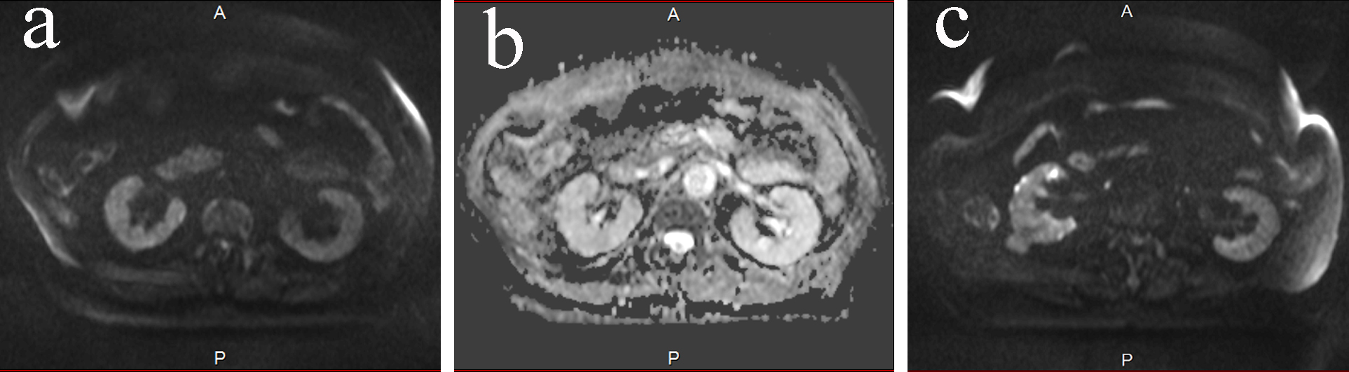
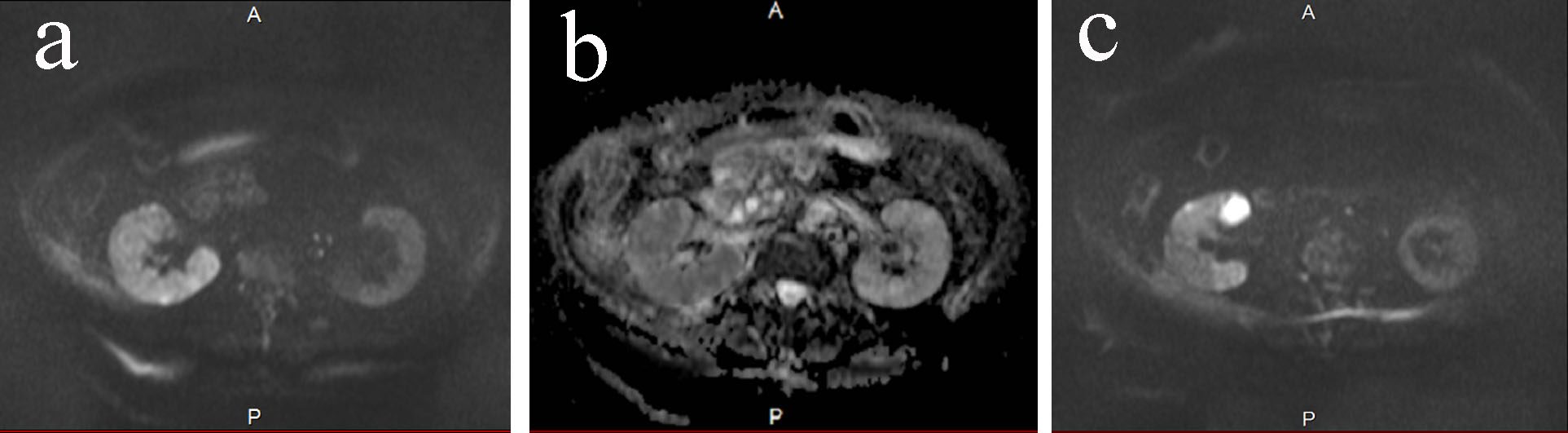
Figure 1. (a) DWMRI on day 0. The high signal intensity was seen in the right kidney. The intensity was apparently higher than the left kidney. (b) ADC map on day 0. The intensity was low in the right kidney. (c) DWMRI on day 0. One of the cyst showed high intensity.
Figure 2.
Figure 2. (a) DWMRI on day 58. The signal intensity in the right kidney lowered, which was obvious comparing to Figure 1a. The intensity was same between the right and the left kidney. (b) ADC map on day 58. Right and left kidney had the same intensity. (c) DWMRI on day 58. The intensity of the infected cyst had decreased.