World Journal of Nephrology and Urology, ISSN 1927-1239 print, 1927-1247 online, Open Access
Article copyright, the authors; Journal compilation copyright, World J Nephrol Urol and Elmer Press Inc
Journal website http://www.wjnu.org

Case Report

Volume 3, Number 3, September 2014, pages 124-128


Hidden Tiger: An Atypical Presentation of Anti-Glomerular Basement Membrane Disease

Figures

Figure 1.
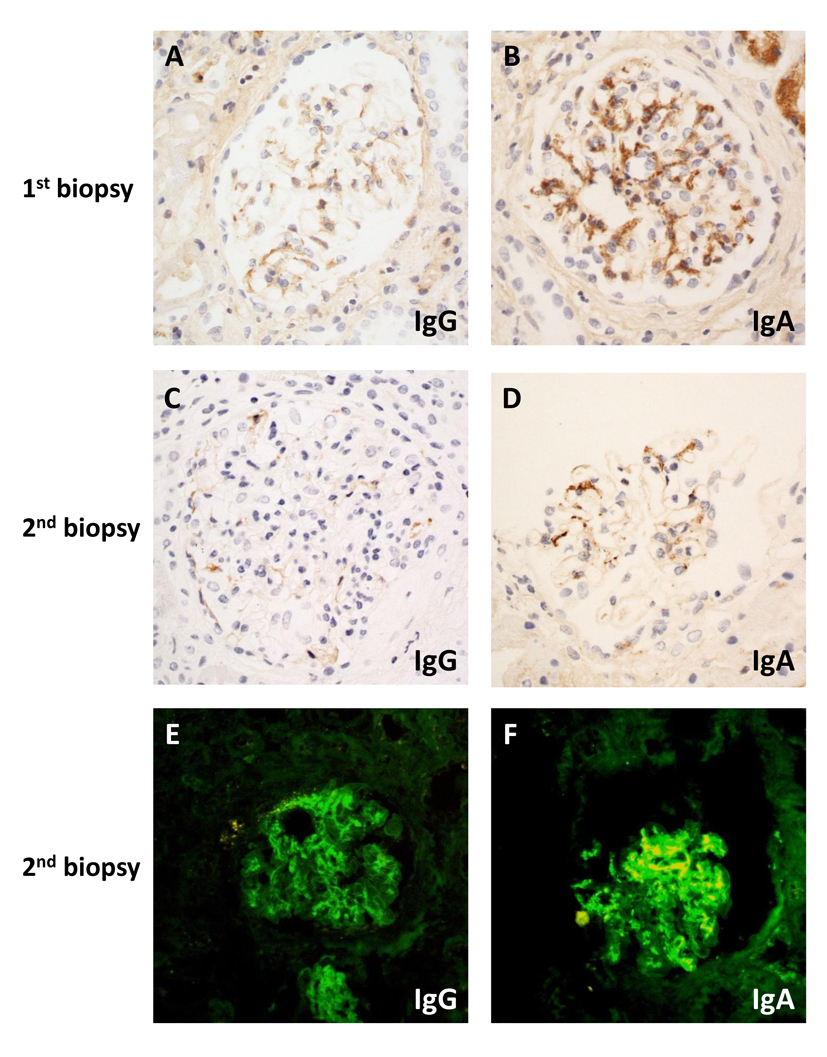
Figure 1. Microscopy of immunostaining on first (A, B) and second (C-F) biopsy by immunohistochemistry (A-D) or direct immunofluorescence (E, F): (A) weak granular mesangial staining for IgG; (B) moderate intensity mesangial staining for IgA; (C) absent IgG; (D) weak mesangial IgA staining; (E) weak linear IgG; (F) strong granular mesangial IgA staining. Original magnification (A-F) × 400.
Figure 2.
Figure 2. Light (A) and electron (B-D) microscopic findings on second biopsy: (A) segmental necrotizing lesion and cellular crescent (silver masson trichrome stain); (B) cellular crescent (× 1,500); (C) defect in glomerular basement membrane (arrowed) (× 4,000); (D) foot process effacement (arrowed) (× 2,500). Original magnification (A) × 400; (B) × 1,500; (C) × 4,000; (D) × 2,500.