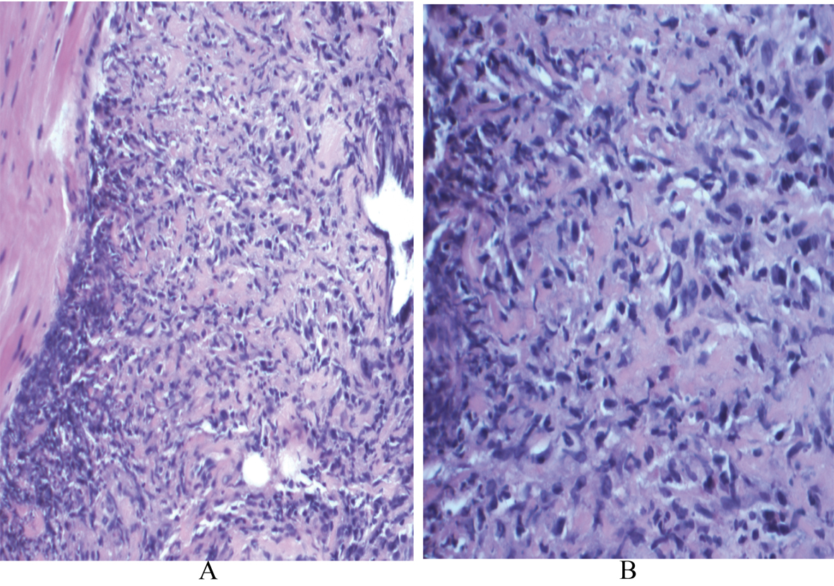
Figure 1. A: Scanning magnification of the prostate tissue shows extensive replacement by granulomatous inflammation (x 1000; H and E); B: High power view demonstrates small foci of necrosis within the granulomas (x 4000; H and E).
| World Journal of Nephrology and Urology, ISSN 1927-1239 print, 1927-1247 online, Open Access |
| Article copyright, the authors; Journal compilation copyright, World J Nephrol Urol and Elmer Press Inc |
| Journal website http://www.wjnu.org |
Case Report
Volume 1, Number 2-3, June 2012, pages 94-97
Granulomatosis With Polyangiitis Presenting as Prostatitis: An Uncommon Scenario
Figures