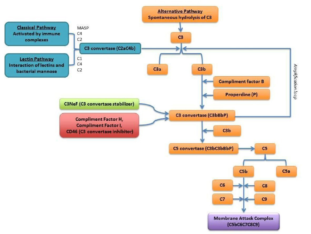
Figure 1. Scheme of the complement system: alternative complement pathway is outlined in orange [8].
| World Journal of Nephrology and Urology, ISSN 1927-1239 print, 1927-1247 online, Open Access |
| Article copyright, the authors; Journal compilation copyright, World J Nephrol Urol and Elmer Press Inc |
| Journal website http://www.wjnu.org |
Case Report
Volume 7, Number 3-4, November 2018, pages 73-77
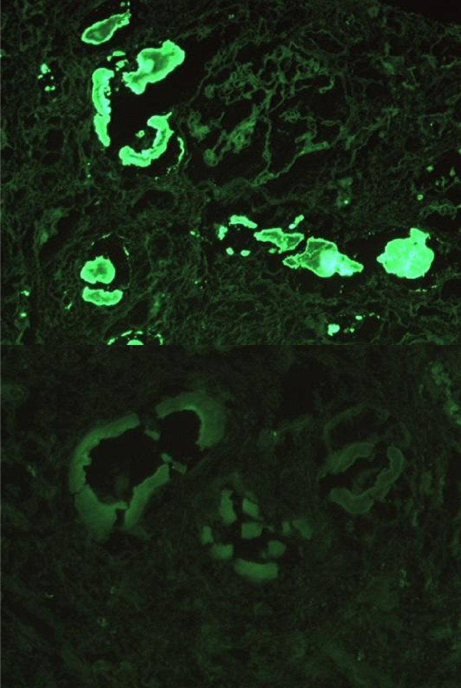
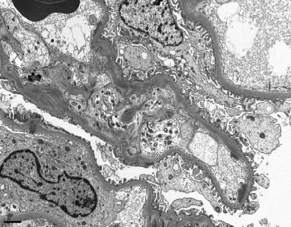
A Case of Myeloma Kidney With Glomerular C3 Deposition
Figures