Figures
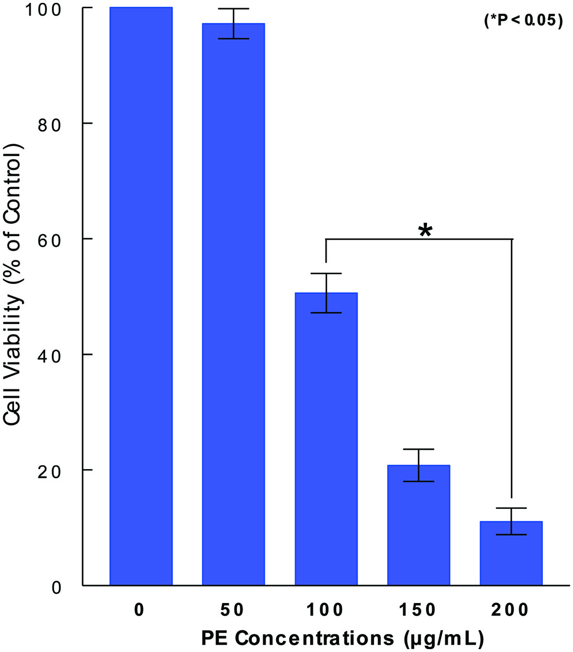
Figure 1. Dose-dependent effect of PE on ACHN cell viability. ACHN cells were cultured with varying concentrations of PE (0 - 200 µg/mL) and cell viability was assessed in 72 h by MTT assay. Cell viability was expressed by the percent (%) of viable cells relative to controls (100%). The data are mean ± standard deviation (SD) from three separate experiments (*P < 0.05 compared with control). PE: Poria mushroom extract; MTT: 3-(4, 5-dimethylthiazol-2-yl)-2, 5-diphenyl tetrazolium bromide.
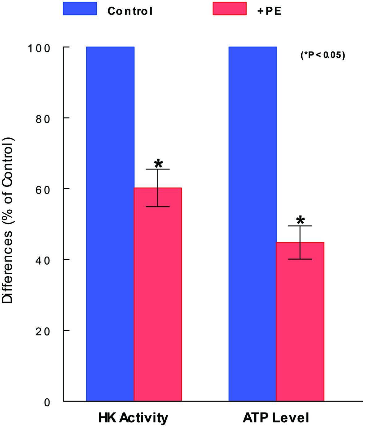
Figure 2. Inhibitory effects of PE on glycolytic parameters. After cells were treated with PE (100 µg/mL) for 72 h and subjected to HK and ATP assays separately. HK activity and ATP level are expressed by the % relative to respective controls (100%). The data are mean ± standard deviation (SD) from three separate experiments (*P < 0.05 compared with control). PE: Poria mushroom extract; HK: hexokinase; ATP: adenosine triphosphate.
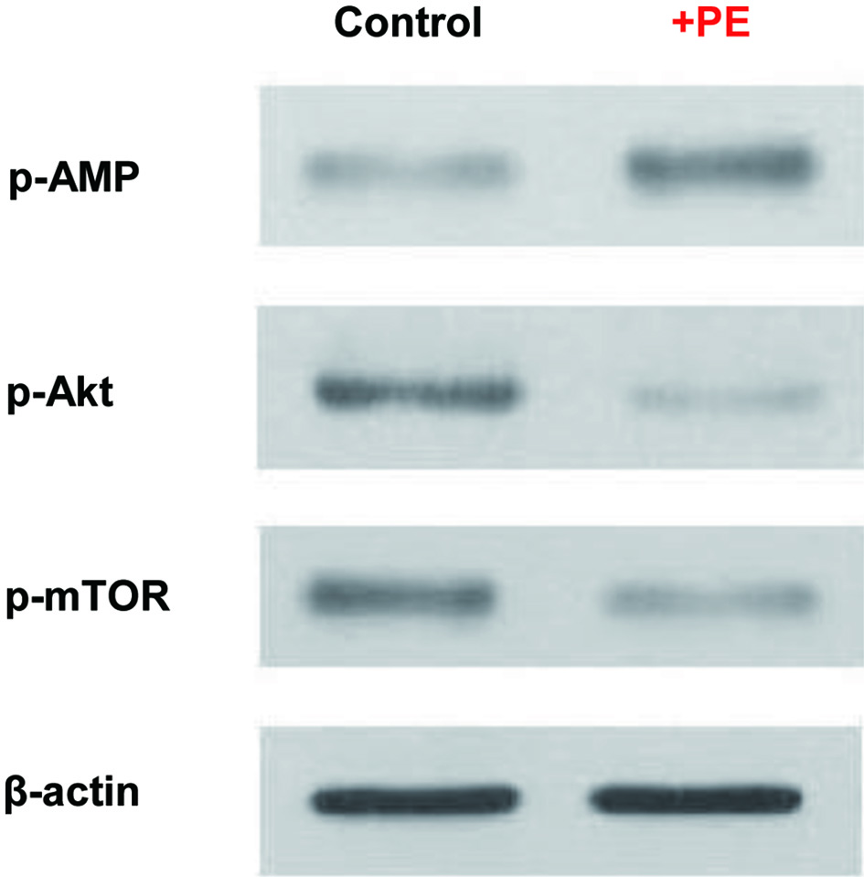
Figure 3. Modulations of metabolic regulators by PE. Cells treated with PE (100 µg/mL) for 72 h were analyzed for four key metabolic regulators using Western blots. Autoradiographs of p-AMPK, p-Akt, and p-mTOR expressed in control and PE-treated cells are shown for comparison. Beta-actin is also shown as a loading control. PE: Poria mushroom extract; AMPK: AMP-activated protein kinase; Akt: protein kinase B; mTOR: mammalian target of rapamycin.
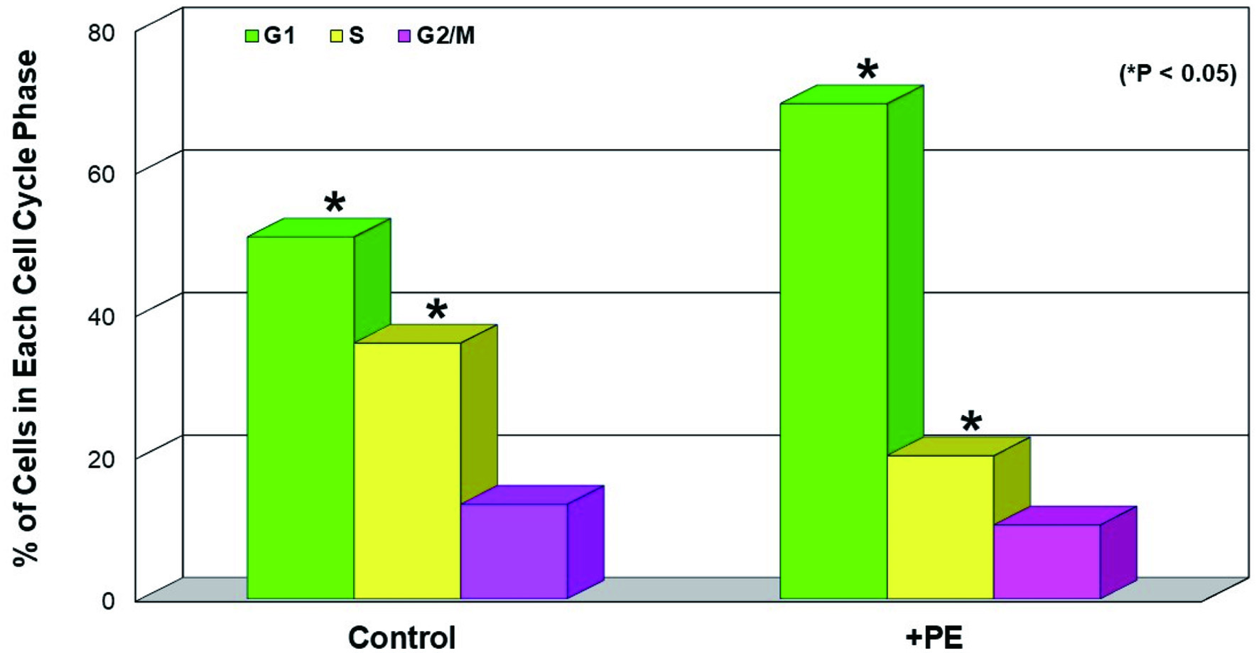
Figure 4. Induction of G1 cell cycle arrest by PE. Cells treated with PE (100 µg/mL) for 72 h were subjected to cell cycle analysis and the % of cell distribution at each cell cycle phase in control and PE-treated cells was plotted. The data are mean ± SD from three separate experiments but only mean values are shown (*P < 0.05 compared with control). PE: Poria mushroom extract; SD: standard deviation.
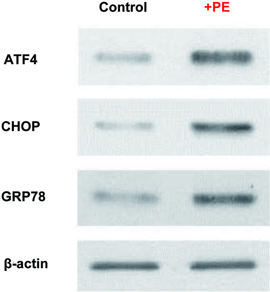
Figure 5. PE induced ER stress. Cells treated with PE (100 µg/mL) for 72 h were analyzed for three ER-related factors, ATF4, CHOP, and GRP78, using Western blots. Autoradiographs of these three factors expressed in control and PE-treated cells are shown for comparison. Beta-actin is also shown as a loading control. PE: Poria mushroom extract; ATF4: activating transcription factor 4; CHOP: C/EBP homologous protein; GRP78: glucose-regulated protein 78.
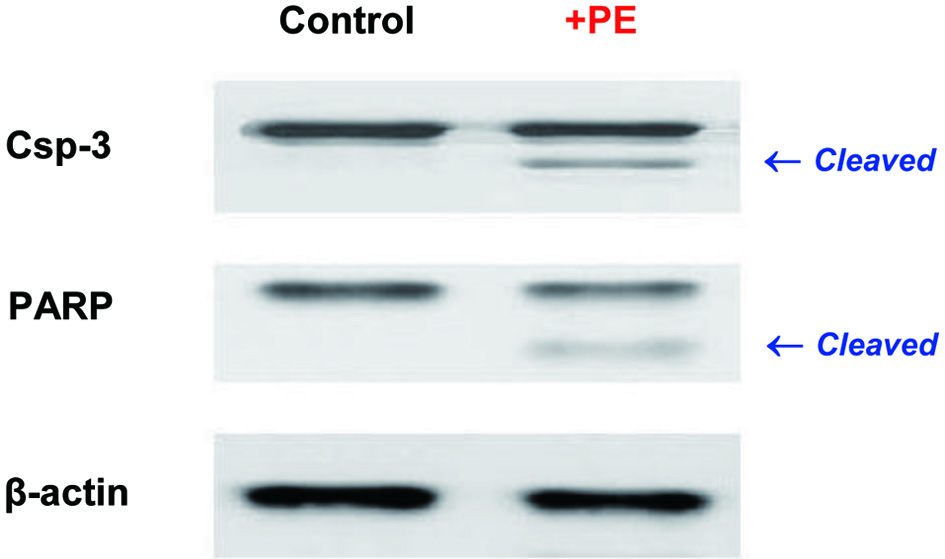
Figure 6. Induction of apoptosis. Cells treated with PE (100 µg/mL) for 72 h were analyzed for two apoptotic regulators using Western blots. Autoradiographs of Csp-3 and PARP in control and PE-treated cells are shown for comparison. The small, cleaved bands by proteolysis in PE-treated cells are indicative of active forms. Beta-actin is also shown as an internal control. PE: Poria mushroom extract; Csp-3: caspace-3; PARP: poly-ADP ribose polymerase.