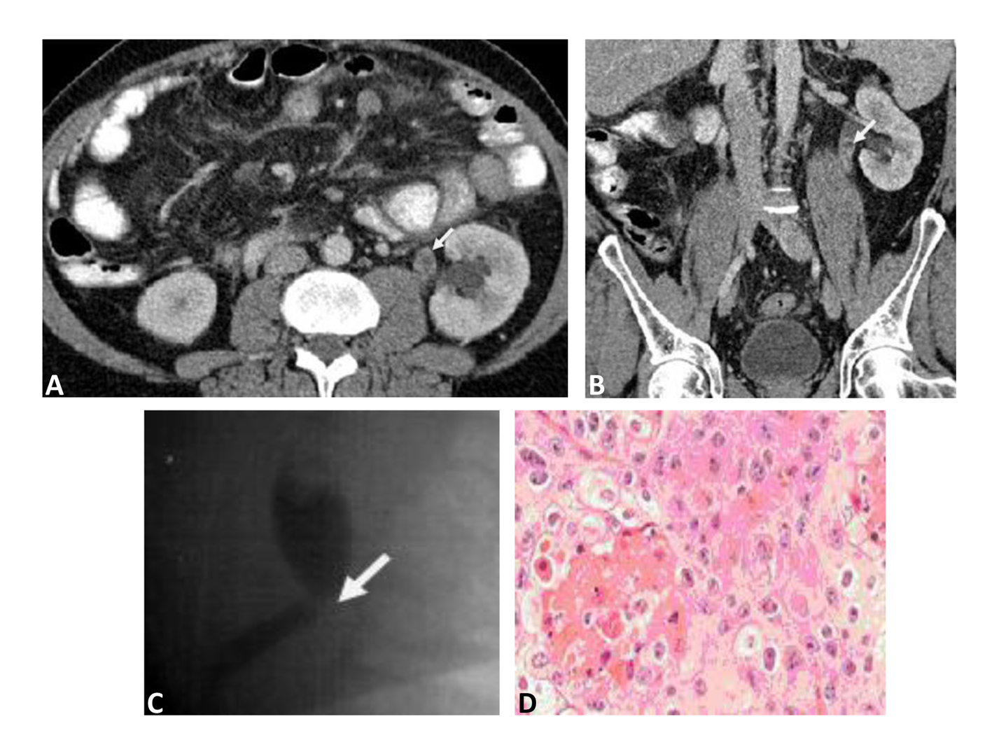
Figure 1. A: Transverse section Contrast Computerized Tomography (CT) image, a filling defect is demonstrated in the left upper ureter (just below the uretero-pelvic junction) (marked by white arrow). B: Sagittal section Contrast Computerized Tomography (CT) image, a filling defect is demonstrated in the left upper ureter (just below the uretero-pelvic junction) (marked by white arrow). C: Retrograde contrast study of left ureter, a filing defect is shown just below the uretero-pelvic junction (marked by white arrow). D: A high power (20 × objectives) of squamous cell carcinoma of the left ureter, nests of squamous cells with hyperchromatic nuclei and prominent keratin production (Haematoxylin and Eosin stain).