World Journal of Nephrology and Urology, ISSN 1927-1239 print, 1927-1247 online, Open Access
Article copyright, the authors; Journal compilation copyright, World J Nephrol Urol and Elmer Press Inc
Journal website http://www.wjnu.org

Case Report

Volume 2, Number 1, June 2013, pages 15-17


Right Testicular Seminoma in Bilateral Cryptorchidism: A Case Report

Figures

Figure 1.
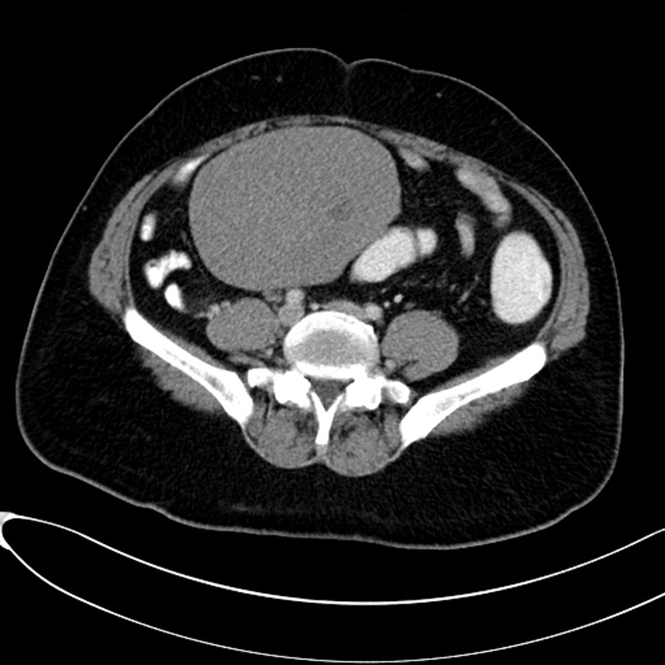
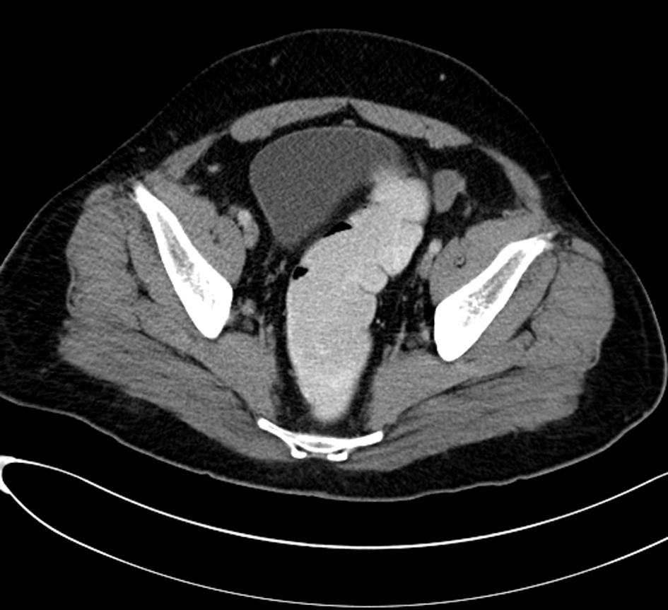
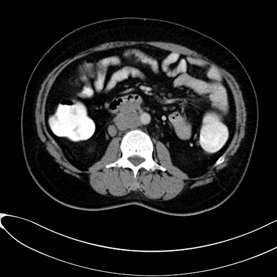
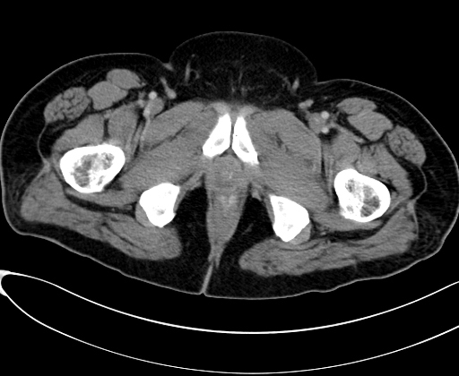
Figure 1. Computed tomography (CT) scan examination shows an empty scrotal sac with absence of the testicles and spermatic cord bilaterally.
Figure 2.
Figure 2. A large homogenous mass is identified within the retro peritoneum of the right lower abdominal quadrant associated with small central area of low attenuation could represent an areas of cystic necrosis. No associated calcification is seen.
Figure 3.
Figure 3. Small oval and homogenous structure is seen within the left aspect of the hemi pelvis just adjacent to the inguinal ring which represents a small atrophied left testicle with no masses.
Figure 4.
Figure 4. There are multiple homogenous and matted lymph nodes metastasis located within the right Para-aortic region in close relation to the right renal hilum.