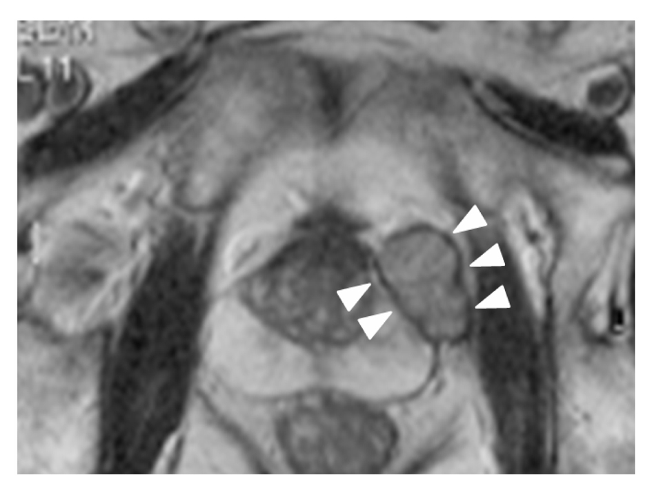
Figure 1. T2-weighted MRI, axial plane. A small, round encapsulated mass is apparent above the prostate. The mass is distinguishable from the prostate.
| World Journal of Nephrology and Urology, ISSN 1927-1239 print, 1927-1247 online, Open Access |
| Article copyright, the authors; Journal compilation copyright, World J Nephrol Urol and Elmer Press Inc |
| Journal website http://www.wjnu.org |
Case Report
Volume 2, Number 1, June 2013, pages 18-20
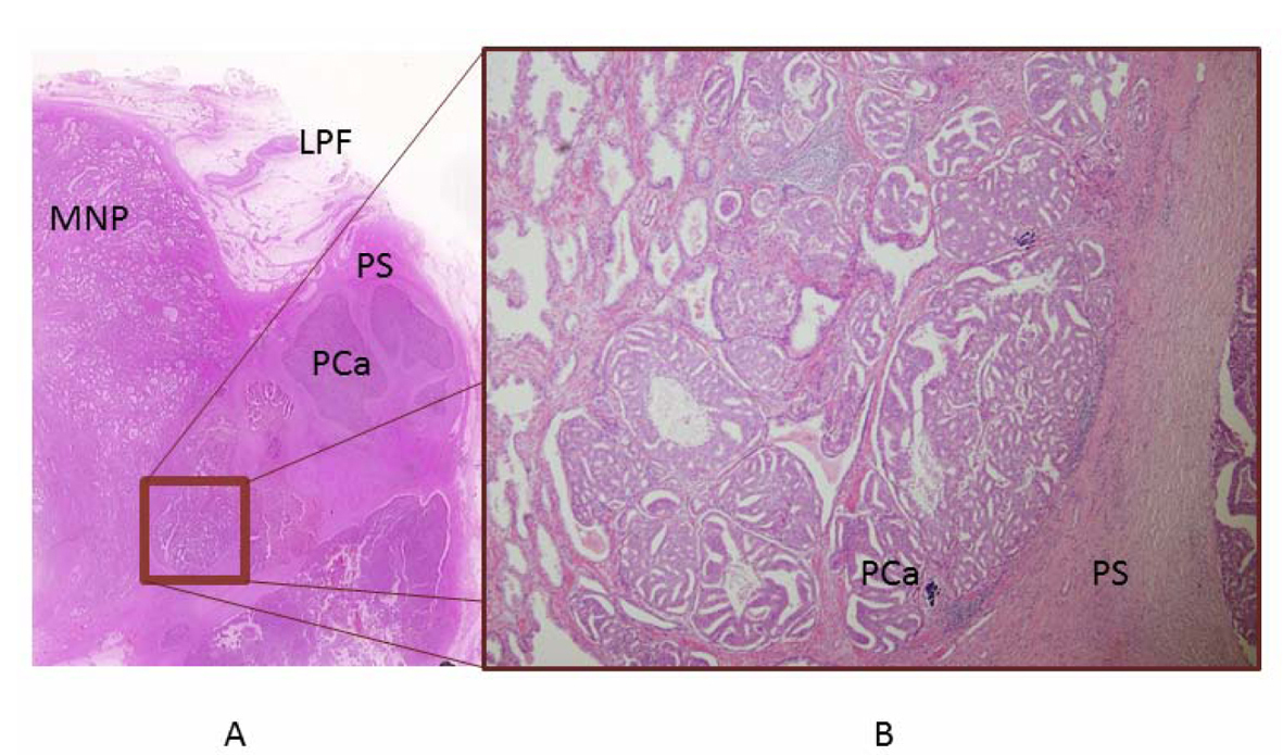
Prostatic Adenocarcinoma With Pseudocapsule
Figures