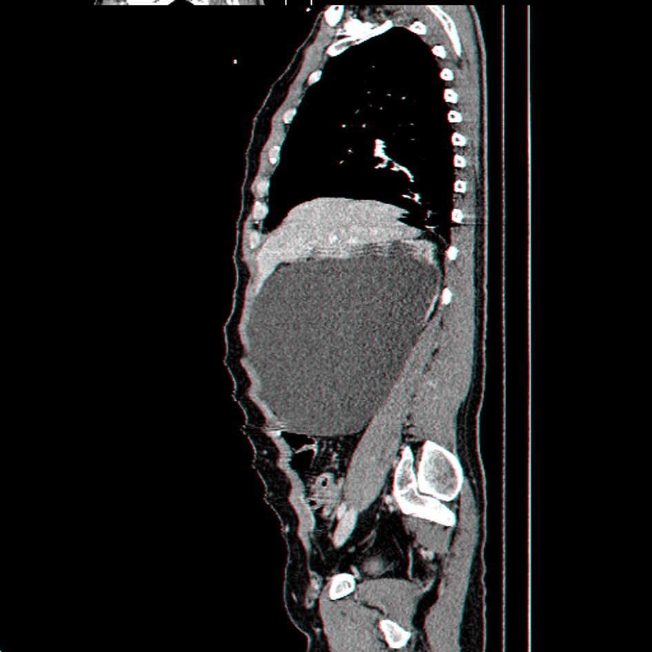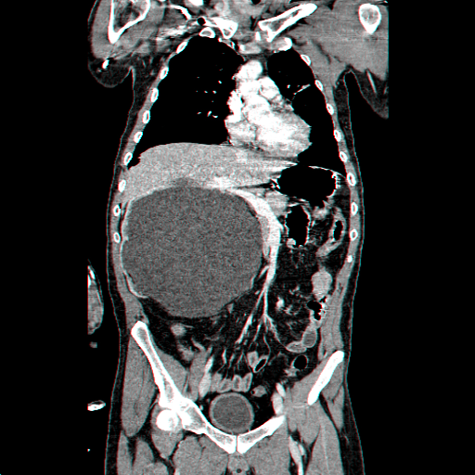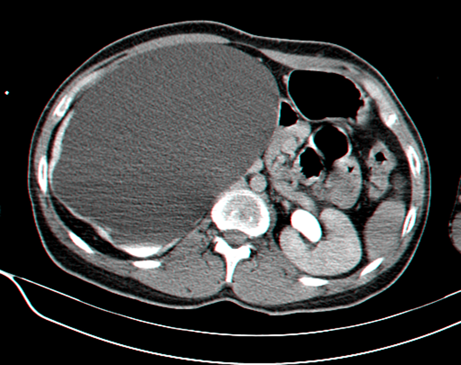
Figure 1. Abdominal CT scan showing giant right hydronephrosis with pressure effect over the bowels and thinning of renal parenchyma.
| World Journal of Nephrology and Urology, ISSN 1927-1239 print, 1927-1247 online, Open Access |
| Article copyright, the authors; Journal compilation copyright, World J Nephrol Urol and Elmer Press Inc |
| Journal website http://www.wjnu.org |
Case Report
Volume 2, Number 1, June 2013, pages 33-35
Giant Hydronephrosis - A Late Diagnosis of Ureteropelvic Junction Obstruction
Figures